
10.7 Medical Applications and Biological Effects of Nuclear Radiation
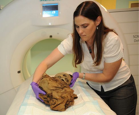
Nuclear physics is an integral part of our everyday lives (Figure 10.26). Radioactive compounds are used to identify cancer, study ancient artifacts, and power our cities. Nuclear fusion also powers the Sun, the primary source of energy on Earth. The focus of this chapter is nuclear radiation. In this section, we ask such questions as: How is nuclear radiation used to benefit society? What are its health risks? How much nuclear radiation is the average person exposed to in a lifetime?
Medical Applications
Most medical and related applications of nuclear physics are driven, at their core, by the difference between a radioactive substance and a non-radioactive substance. One of the first such methods is the precision measurement and detection method known as radioimmunoassay (RIA). Developed by Rosalyn Sussman Yalow and Solomon Berson in the late 1950s, RIA relies on the principle of competitive binding. For the particular substance being measured, a sample containing a radioactive isotope is prepared. A known quantity of antibodies is then introduced. By measuring the amount of "unbound" antibodies after the reaction, technicians can detect and measure the precise amount of the target substance. Radioimmunoassay is essential in cancer screening, hepatitus diagnosis, narcotics investigation, and other analyses.
One of the most important uses of nuclear radiation is the location and study of diseased tissue. This application requires a special drug called a radiopharmaceutical. A radiopharmaceutical contains an unstable radioactive isotope. When the drug enters the body, it tends to concentrate in inflamed regions of the body. (Recall that the interaction of the drug with the body does not depend on whether a given nucleus is replaced by one of its isotopes, since this interaction is determined by chemical interactions.) Radiation detectors used outside the body use nuclear radiation from the radioisotopes to locate the diseased tissue. Radiopharmaceuticals are called radioactive tags because they allow doctors to track the movement of drugs in the body. Radioactive tags are for many purposes, including the identification of cancer cells in the bones, brain tumors, and Alzheimer’s disease (Figure 10.27). Radioactive tags are also used to monitor the function of body organs, such as blood flow, heart muscle activity, and iodine uptake in the thyroid gland.
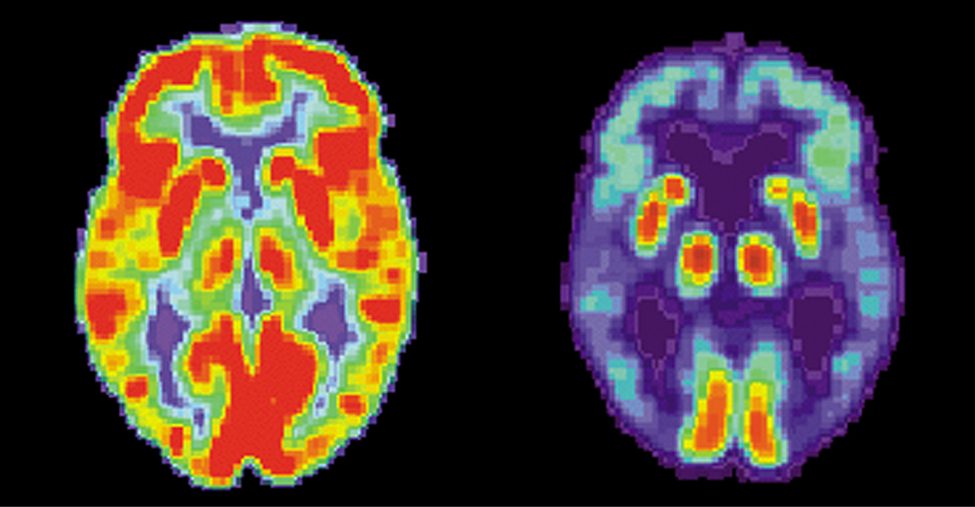
Table 10.2 lists some medical diagnostic uses of radiopharmaceuticals, including isotopes and typical activity (A) levels. One common diagnostic test uses iodine to image the thyroid, since iodine is concentrated in that organ. Another common nuclear diagnostic is the thallium scan for the cardiovascular system, which reveals blockages in the coronary arteries and examines heart activity. The salt TlCl can be used because it acts like NaCl and follows the blood. Note that Table 10.2 lists many diagnostic uses for , where “m” stands for a metastable state of the technetium nucleus. This isotope is used in many compounds to image the skeleton, heart, lungs, and kidneys. About of all radiopharmaceuticals employ because it produces a single, easily identified, 0.142-MeV ray and has a short 6.0-h half-life, which reduces radiation exposure.
| Procedure, Isotope | Activity (mCi), where | Procedure, Isotope | Activity (mCi), where |
|---|---|---|---|
| Brain scan | Thyroid scan | ||
| 7.5 | 0.05 | ||
| (PET) | 50 | 0.07 | |
| Lung scan | Liver scan | ||
| 7.5 | (colloid) | 0.1 | |
| 2 | (colloid) | 2 | |
| Cardiovascular blood pool | Bone scan | ||
| 0.2 | 0.1 | ||
| 2 | 10 | ||
| Cardiovascular arterial flow | Kidney scan | ||
| 3 | 0.1 | ||
| 7.5 | 1.5 | ||
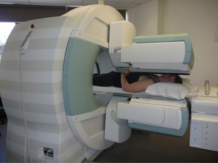
The first radiation detectors produced two-dimensional images, like a photo taken from a camera. However, a circular array of detectors that can be rotated can be used to produce three-dimensional images. This technique is similar to that used in X-ray computed tomography (CT) scans. One application of this technique is called single-photon-emission CT (SPECT) (Figure 10.28). The spatial resolution of this technique is about 1 cm.
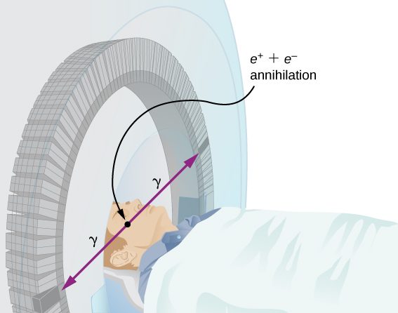
Improved image resolution is achieved by a technique known as positron emission tomography (PET). This technique use radioisotopes that decay by radiation. When a positron encounters an electron, these particle annihilate to produce two gamma-ray photons. This reaction is represented by
These -ray photons have identical 0.511-MeV energies and move directly away from one another (Figure 10.29). This easily identified decay signature can be used to identify the location of the radioactive isotope. Examples of -emitting isotopes used in PET include . The nuclei have the advantage of being able to function as tags for natural body compounds. Its resolution of 0.5 cm is better than that of SPECT.
PET scans are especially useful to examine the brain’s anatomy and function. For example, PET scans can be used to monitor the brain’s use of oxygen and water, identify regions of decreased metabolism (linked to Alzheimer’s disease), and locate different parts of the brain responsible for sight, speech, and fine motor activity
Biological Effects
Nuclear radiation can have both positive and negative effects on biological systems. However, it can also be used to treat and even cure cancer. How do we understand these effects? To answer this question, consider molecules within cells, particularly DNA molecules.
Cells have long, double-helical DNA molecules containing chemical codes that govern the function and processes of the cell. Nuclear radiation can alter the structural features of the DNA chain, leading to changes in the genetic code. In human cells, we can have as many as a million individual instances of damage to DNA per cell per day. DNA contains codes that check whether the DNA is damaged and can repair itself. This repair ability of DNA is vital for maintaining the integrity of the genetic code and for the normal functioning of the entire organism. It should be constantly active and needs to respond rapidly. The rate of DNA repair depends on various factors such as the type and age of the cell. If nuclear radiation damages the ability of the cell to repair DNA, the cell can
- Retreat to an irreversible state of dormancy (known as senescence);
- Commit suicide (known as programmed cell death); or
- Progress into unregulated cell division, possibly leading to tumors and cancers.
Nuclear radiation can harm the human body is many other ways as well. For example, high doses of nuclear radiation can cause burns and even hair loss.
Biological effects of nuclear radiation are expressed by many different physical quantities and in many different units. A common unit to express the biological effects of nuclear radiation is the rad or radiation dose unit. One rad is equal to 1/100 of a joule of nuclear energy deposited per kilogram of tissue, written:
For example, if a 50.0-kg person is exposed to nuclear radiation over their entire body and absorbs 1.00 J, then their whole-body radiation dose is
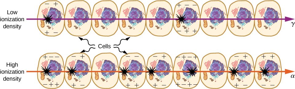
Nuclear radiation damages cells by ionizing atoms in the cells as they pass through the cells (Figure 10.30). The effects of ionizing radiation depend on the dose in rads, but also on the type of radiation (alpha, beta, gamma, or X-ray) and the type of tissue. For example, if the range of the radiation is small, as it is for rays, then the ionization and the damage created is more concentrated and harder for the organism to repair. To account for such affects, we define the relative biological effectiveness (RBE). Sample RBE values for several types of ionizing nuclear radiation are given in Table 10.3.
| Type and Energy of Radiation | RBE[1] |
|---|---|
| X-rays | 1 |
| rays | 1 |
| rays greater than 32 keV | 1 |
| rays less than 32 keV | 1.7 |
| Neutrons, thermal to slow (<20 keV) | 2–5 |
| Neutrons, fast (1–10 MeV) | 10 (body), 32 (eyes) |
| Protons (1–10 MeV) | 10 (body), 32 (eyes) |
| rays from radioactive decay | 10–20 |
| Heavy ions from accelerators | 10–20 |
A dose unit more closely related to effects in biological tissue is called the roentgen equivalent man (rem) and is defined to be the dose (in rads) multiplied by the relative biological effectiveness (RBE). Thus, if a person had a whole-body dose of 2.00 rad of radiation, the dose in rem would be rem for the whole body. If the person had a whole-body dose of 2.00 rad of radiation, then the dose in rem would be rem for the whole body. The rays would have 20 times the effect on the person than the rays for the same deposited energy. The SI equivalent of the rem, and the more standard term, is the sievert (Sv) is
The RBEs given in Table 10.3 are approximate but reflect an understanding of nuclear radiation and its interaction with living tissue. For example, neutrons are known to cause more damage than rays, although both are neutral and have large ranges, due to secondary radiation. Any dose less than 100 mSv (10 rem) is called a low dose, 0.1 Sv to 1 Sv (10 to 100 rem) is called a moderate dose, and anything greater than 1 Sv (100 rem) is called a high dose. It is difficult to determine if a person has been exposed to less than 10 mSv.
Biological effects of different levels of nuclear radiation on the human body are given in Table 10.4. The first clue that a person has been exposed to radiation is a change in blood count, which is not surprising since blood cells are the most rapidly reproducing cells in the body. At higher doses, nausea and hair loss are observed, which may be due to interference with cell reproduction. Cells in the lining of the digestive system also rapidly reproduce, and their destruction causes nausea. When the growth of hair cells slows, the hair follicles become thin and break off. High doses cause significant cell death in all systems, but the lowest doses that cause fatalities do so by weakening the immune system through the loss of white blood cells.
| Dose in Sv[1] | Effect |
|---|---|
| 0–0.10 | No observable effect. |
| 0.1–1 | Slight to moderate decrease in white blood cell counts. |
| 0.5 | Temporary sterility; 0.35 for women, 0.50 for men. |
| 1–2 | Significant reduction in blood cell counts, brief nausea and vomiting. Rarely fatal. |
| 2–5 | Nausea, vomiting, hair loss, severe blood damage, hemorrhage, fatalities. |
| 4.5 | Lethal to of the population within 32 days after exposure if not treated. |
| 5–20 | Worst effects due to malfunction of small intestine and blood systems. Limited survival. |
| >20 | Fatal within hours due to collapse of central nervous system. |
Sources of Radiation
Human are also exposed to many sources of nuclear radiation. A summary of average radiation doses for different sources by country is given in Table 10.5. Earth emits radiation due to the isotopes of uranium, thorium, and potassium. Radiation levels from these sources depend on location and can vary by a factor of 10. Fertilizers contain isotopes of potassium and uranium, which we digest in the food we eat. Fertilizers have more than 3000 Bq/kg radioactivity, compared to just 66 Bq/kg for Carbon-14.
| Source | Dose (mSv/y)[1] | |||
|---|---|---|---|---|
| Australia | Germany | US | World | |
| Natural radiation – external | ||||
| Cosmic rays | 0.30 | 0.28 | 0.30 | 0.39 |
| Soil, building materials | 0.40 | 0.40 | 0.30 | 0.48 |
| Radon gas | 0.90 | 1.1 | 2.0 | 1.2 |
| Natural radiation – internal | ||||
| 0.24 | 0.28 | 0.40 | 0.29 | |
| Artificial radiation | ||||
| Medical and dental | 0.80 | 0.90 | 0.53 | 0.40 |
| TOTAL | 2.6 | 3.0 | 3.5 | 2.8 |
Medical visits are also a source of nuclear radiation. A sample of common nuclear radiation doses is given in Table 10.6. These doses are generally low and can be lowered further with improved techniques and more sensitive detectors. With the possible exception of routine dental X-rays, medical use of nuclear radiation is used only when the risk-benefit is favorable. Chest X-rays give the lowest doses—about 0.1 mSv to the tissue affected, with less than scattering into tissues that are not directly imaged. Other X-ray procedures range upward to about 10 mSv in a CT scan, and about 5 mSv (0.5 rem) per dental X-ray, again both only affecting the tissue imaged. Medical images with radiopharmaceuticals give doses ranging from 1 to 5 mSv, usually localized.
| Procedure | Effective Dose (mSv) |
|---|---|
| Chest | 0.02 |
| Dental | 0.01 |
| Skull | 0.07 |
| Leg | 0.02 |
| Mammogram | 0.40 |
| Barium enema | 7.0 |
| Upper GI | 3.0 |
| CT head | 2.0 |
| CT abdomen | 10.0 |
Summary
- Nuclear technology is used in medicine to locate and study diseased tissue using special drugs called radiopharmaceuticals. Radioactive tags are used to identify cancer cells in the bones, brain tumors, and Alzheimer’s disease, and to monitor the function of body organs, such as blood flow, heart muscle activity, and iodine uptake in the thyroid gland.
- The biological effects of ionizing radiation are due to two effects it has on cells: interference with cell reproduction and destruction of cell function.
- Common sources of radiation include that emitted by Earth due to the isotopes of uranium, thorium, and potassium; natural radiation from cosmic rays, soils, and building materials, and artificial sources from medical and dental diagnostic tests.
- Biological effects of nuclear radiation are expressed by many different physical quantities and in many different units, including the rad or radiation dose unit.
Key Equations
| Atomic mass number | |
|---|---|
| Standard format for expressing an isotope | |
| Nuclear radius, where r0 is the radius of a single proton | |
| Mass defect | |
| Binding energy | |
| Binding energy per nucleon | |
| Radioactive decay rate | |
| Radioactive decay law | |
| Decay constant | |
| Lifetime of a substance | |
| Activity of a radioactive substance | |
| Activity of a radioactive substance (linear form) | |
| Alpha decay | |
| Beta decay | |
| Positron emission | |
| Gamma decay |
Conceptual Questions
Why is a PET scan more accurate than a SPECT scan?
Isotopes that emit radiation are relatively safe outside the body and exceptionally hazardous inside. Explain why.
Alpha particles do not penetrate materials such as skin and clothes easily. (Recall that alpha radiation is barely able to pass through a thin sheet of paper.) However, when produce inside the body, neighboring cells are vulnerable.
Ionizing radiation can impair the ability of a cell to repair DNA. What are the three ways the cell can respond?
Problems
What is the dose in mSv for: (a) a 0.1-Gy X-ray? (b) 2.5 mGy of neutron exposure to the eye? (c) 1.5m Gy of exposure?
Find the radiation dose in Gy for: (a) A 10-mSv fluoroscopic X-ray series. (b) 50 mSv of skin exposure by an emitter. (c) 160 mSv of and rays from the in your body.
: a. 0.01 Gy; b. 0.0025 Gy; c. 0.16 Gy
Find the mass of that has an activity of .
In the 1980s, the term picowave was used to describe food irradiation in order to overcome public resistance by playing on the well-known safety of microwave radiation. Find the energy in MeV of a photon having a wavelength of a picometer.
1.24 MeV
What is the dose in Sv in a cancer treatment that exposes the patient to 200 Gy of rays?
One half the rays from are absorbed by a 0.170-mm-thick lead shielding. Half of the rays that pass through the first layer of lead are absorbed in a second layer of equal thickness. What thickness of lead will absorb all but one in 1000 of these rays?
1.69 mm
How many Gy of exposure is needed to give a cancerous tumor a dose of 40 Sv if it is exposed to activity?
A plumber at a nuclear power plant receives a whole-body dose of 30 mSv in 15 minutes while repairing a crucial valve. Find the radiation-induced yearly risk of death from cancer and the chance of genetic defect from this maximum allowable exposure.
For cancer: The risk each year of dying from induced cancer is 30 in a million. For genetic defect: The chance each year of an induced genetic defect is 10 in a million.
Calculate the dose in rem/y for the lungs of a weapons plant employee who inhales and retains an activity of in an accident. The mass of affected lung tissue is 2.00 kg and the plutonium decays by emission of a 5.23-MeV particle. Assume a RBE value of 20.
Additional Problems
The wiki-phony site states that the atomic mass of chlorine is 40 g/mol. Check this result. Hint: The two, most common stable isotopes of chlorine are: and . (The abundance of Cl-35 is , and the abundance of Cl-37 is .)
A particle physicist discovers a neutral particle with a mass of 2.02733 u that he assumes is two neutrons bound together.
(a) Find the binding energy.
(b) What is unreasonable about this result?
A nuclear physicist finds of in a piece of uranium ore (T1/2 = ). (a) Use the decay law to determine how much would had to have been on Earth when it formed ago for to be left today. (b) What is unreasonable about this result? (c) How is this unreasonable result resolved?
a. ; b. This mass is impossibly large; it is greater than the mass of the entire Milky Way galaxy. c. is not produced through natural processes operating over long times on Earth, but through artificial processes in a nuclear reactor.
A group of scientists use carbon dating to date a piece of wood to be 3 billion years old. Why doesn’t this make sense?
According to your lab partner, a 2.00-cm-thick sodium-iodide crystal absorbs all but of rays from a radioactive source and a 4.00-cm piece of the same material absorbs all but Is this result reasonable?
If of rays are left after 2.00 cm, then only are left after 4.00 cm. This is much smaller than your lab partner’s result ().
In the science section of the newspaper, an article reports the efforts of a group of scientists to create a new nuclear reactor based on the fission of iron (Fe). Is this a good idea?
The ceramic glaze on a red-orange “Fiestaware” plate is and contains 50.0 grams of , but very little . (a) What is the activity of the plate? (b) Calculate the total energy that will be released by the decay. (c) If energy is worth 12.0 cents per , what is the monetary value of the energy emitted? (These brightly-colored ceramic plates went out of production some 30 years ago, but are still available as collectibles.)
a. ; (b) From Appendix B, the energy released per decay is 4.27 MeV, so ; (c) The monetary value of the energy is
Large amounts of depleted uranium are available as a by-product of uranium processing for reactor fuel and weapons. Uranium is very dense and makes good counter weights for aircraft. Suppose you have a 4000-kg block of . (a) Find its activity. (b) How many calories per day are generated by thermalization of the decay energy? (c) Do you think you could detect this as heat? Explain.
A piece of wood from an ancient Egyptian tomb is tested for its carbon-14 activity. It is found to have an activity per gram of carbon of . What is the age of the wood?
We know that and
Thus, the age of the tomb is
Challenge Problems
This problem demonstrates that the binding energy of the electron in the ground state of a hydrogen atom is much smaller than the rest mass energies of the proton and electron.
(a) Calculate the mass equivalent in u of the 13.6-eV binding energy of an electron in a hydrogen atom, and compare this with the known mass of the hydrogen atom.
(b) Subtract the known mass of the proton from the known mass of the hydrogen atom.
(c) Take the ratio of the binding energy of the electron (13.6 eV) to the energy equivalent of the electron’s mass (0.511 MeV).
(d) Discuss how your answers confirm the stated purpose of this problem.
The Galileo space probe was launched on its long journey past Venus and Earth in 1989, with an ultimate goal of Jupiter. Its power source is 11.0 kg of a by-product of nuclear weapons plutonium production. Electrical energy is generated thermoelectrically from the heat produced when the 5.59-MeV particles emitted in each decay crash to a halt inside the plutonium and its shielding. The half-life of is 87.7 years.
(a) What was the original activity of the in becquerels?
(b) What power was emitted in kilowatts?
(c) What power was emitted 12.0 y after launch? You may neglect any extra energy from daughter nuclides and any losses from escaping rays.
a. ; b. 6.24 kW; c. 5.67 kW
Find the energy emitted in the decay of .
Engineers are frequently called on to inspect and, if necessary, repair equipment in nuclear power plants. Suppose that the city lights go out. After inspecting the nuclear reactor, you find a leaky pipe that leads from the steam generator to turbine chamber. (a) How do the pressure readings for the turbine chamber and steam condenser compare? (b) Why is the nuclear reactor not generating electricity?
a. Due to the leak, the pressure in the turbine chamber has dropped significantly. The pressure difference between the turbine chamber and steam condenser is now very low. b. A large pressure difference is required for steam to pass through the turbine chamber and turn the turbine.
If two nuclei are to fuse in a nuclear reaction, they must be moving fast enough so that the repulsive Coulomb force between them does not prevent them for getting within of one another. At this distance or nearer, the attractive nuclear force can overcome the Coulomb force, and the nuclei are able to fuse.
(a) Find a simple formula that can be used to estimate the minimum kinetic energy the nuclei must have if they are to fuse. To keep the calculation simple, assume the two nuclei are identical and moving toward one another with the same speed v. (b) Use this minimum kinetic energy to estimate the minimum temperature a gas of the nuclei must have before a significant number of them will undergo fusion. Calculate this minimum temperature first for hydrogen and then for helium. (Hint: For fusion to occur, the minimum kinetic energy when the nuclei are far apart must be equal to the Coulomb potential energy when they are a distance R apart.)
For the reaction, , find the amount of energy transfers to and (on the right side of the equation). Assume the reactants are initially at rest. (Hint: Use conservation of momentum principle.)
The energies are
. Notice that most of the energy goes to the ray.
Engineers are frequently called on to inspect and, if necessary, repair equipment in medical hospitals. Suppose that the PET system malfunctions. After inspecting the unit, you suspect that one of the PET photon detectors is misaligned. To test your theory you position one detector at the location relative to a radioactive test sample at the center of the patient bed. (a) If the second photon detector is properly aligned where should it be located? (b) What energy reading is expected?